Biomedical Imaging
Biomedical Imaging
Biomedical imaging is increasingly becoming popular with rapid upgrades in both traditional and modern imaging systems. Imaging science has applications both in vivo and in vitro, both in real-time and with pre-recorded data. Confocal microscopy, endoscopy, and computed tomography are some common examples. Biomedical images and videos may suffer poor visibility due to a variety of reasons. One example case study could be taken from live 3D confocal microscopy.
Live specimens have a limit on laser exposure which they can endure. Frequent overexposure can easily damage or kill the specimen. Hence, the strength of the laser is often controlled at medium or low value. The incident laser beam rapidly gets absorbed when images corresponding to bottom layer tissues are acquired. This results in low intensity (or low visibility) images for deeper slices.
The problem cannot be overcome by increasing the strength of the laser. So the only solution is to achieve a faithful restoration of weak signals and normalize amplitudes of oscillations corresponding to all slices to have similar sharpness and contrast. This is a task which our technology can easily achieve.
Similarly, other biomedical images can suffer different forms of illumination challenges. Endoscopy of lungs for example may suffer haziness due to the presence of moisture in the lungs, resulting in poor visibility. We are at an early stage of our development work in the area of biomedical images and more research is currently on its way.
Sample: Biomedical Imaging
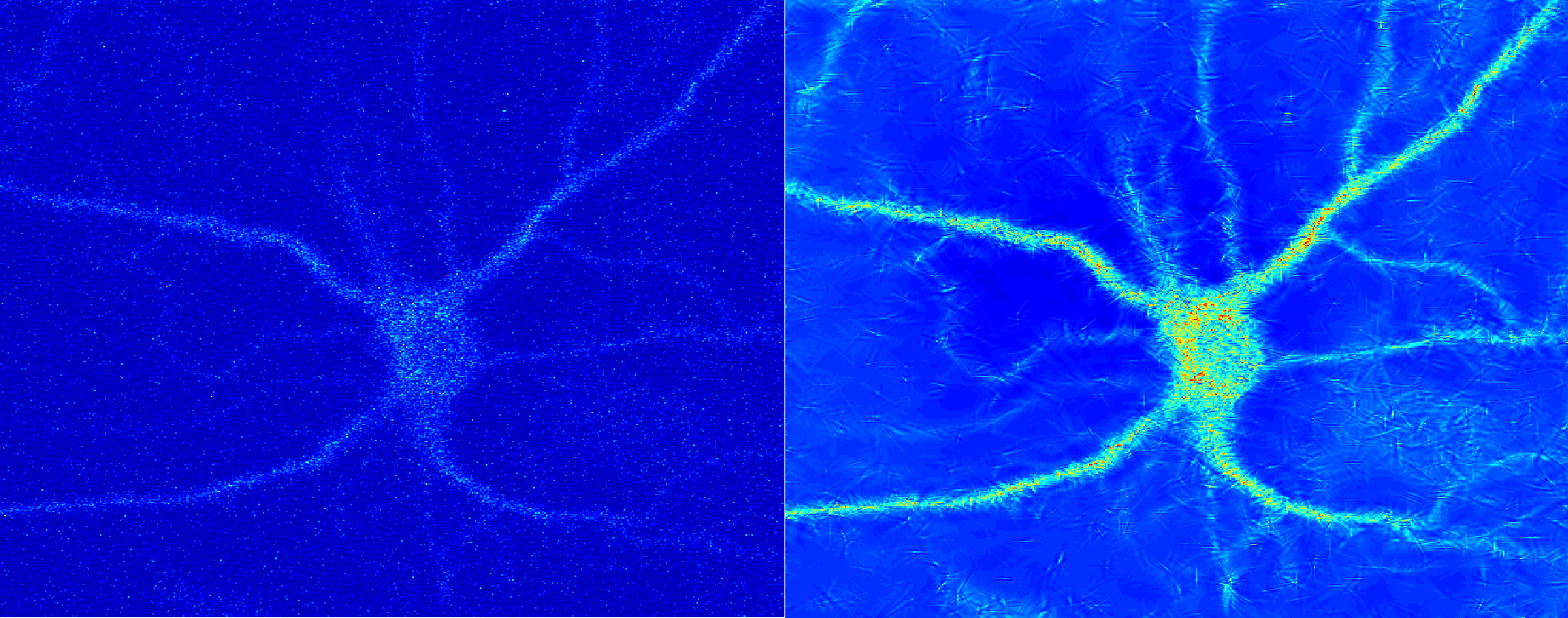
Figure: An example where we enhance the visibility of neuron cells present in the bottom stack of cell culture.